Abberior DyMIN STED
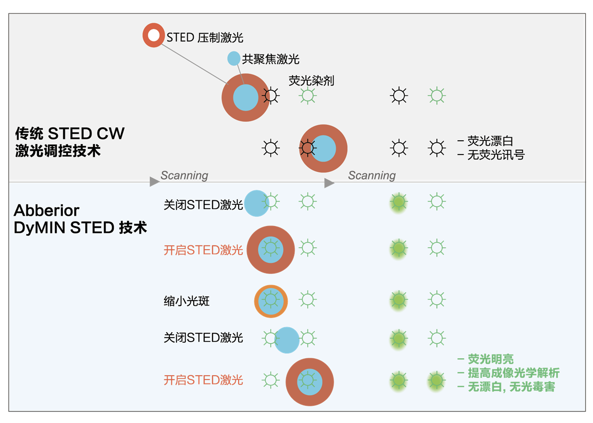
选配的智能光照调控模块精准控制共聚焦激光探寻荧光分子团, 再启动限缩 STED 激光于荧光分子团区域.
在荧光成像, 仅需微小的光剂量, 避免荧光淬灭与光毒害, 提高STED超分辨的光学分辨力
详细介绍

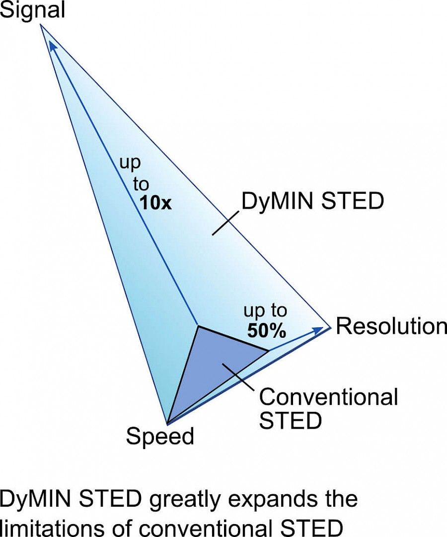
DyMIN STED 智能光照调控
探寻出荧光分子团,找到精准的位置后,再启动该区域的STED激光,将STED激光的使限缩在更小的荧光分子团内,进一步减少了应用最大STED功率的样品区域。所以,更进一步的减少荧光淬灭的发生的发生. 同时再进一步提高 STED 超分辨的光学分辨力. 在长时间活细胞超分辨成像, 纯光学解析可达 25 nm, 可以分辨相距 30 nm 的两个蛋白分子.
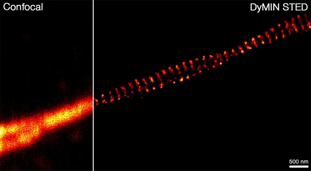
DyMIN STED very clearly resolves spectrin rings with superior signal-to-noise ratio. Primary hippocampal neurons (22 days in vitro) show the characteristic ~192 nm betaII spectrin periodicity along distal axons. Dye: Abberior STAR 635P, Excitation: 635 nm, STED: 775 nm. Sample with courtesy from Elisa D’Este (MPIbpc).
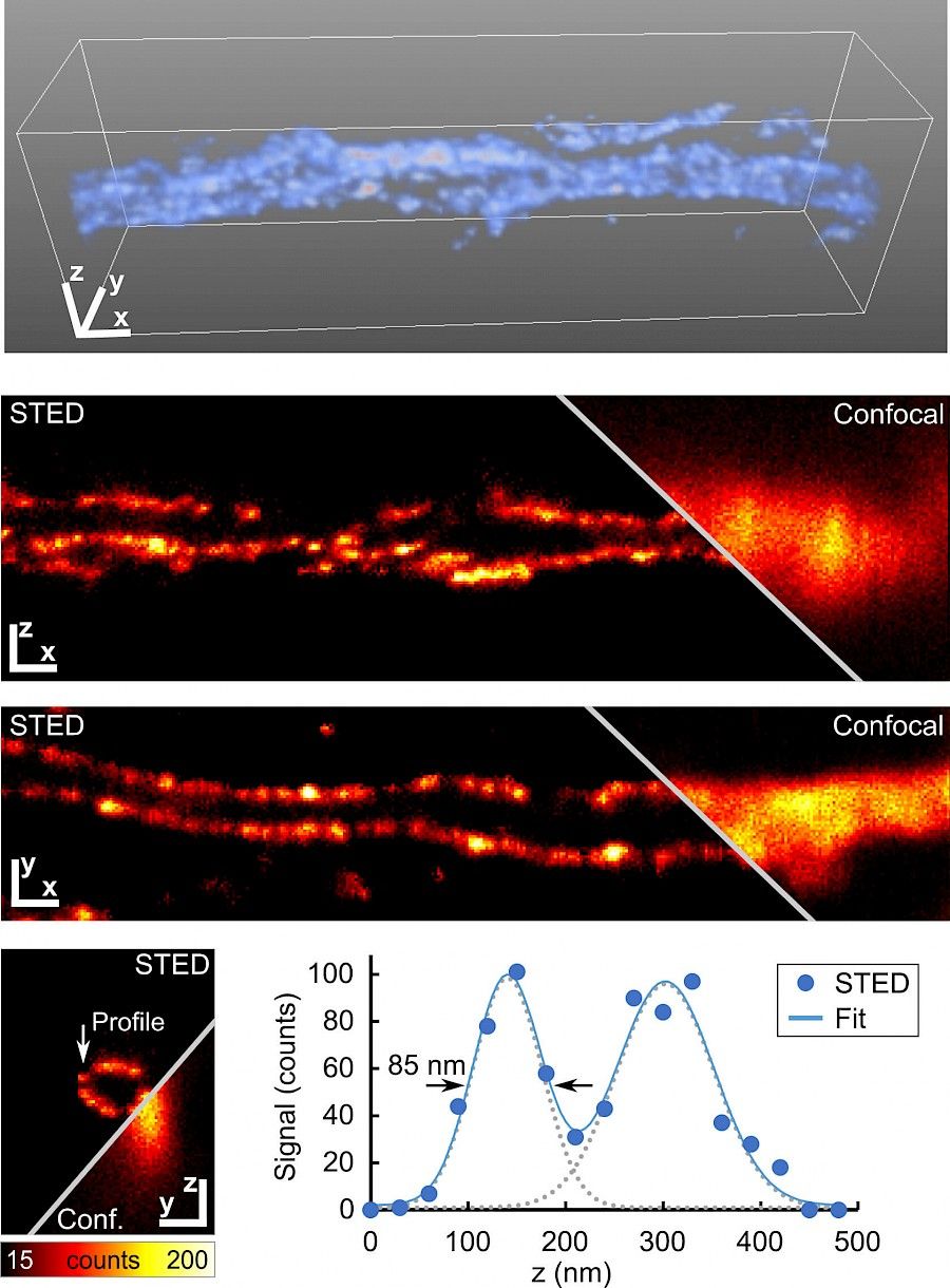
Easy3D DyMIN STED significantly reduces bleaching and thus enables the acquisition of a volume stack with an isotropic resolution of ~85 nm. Primary hippocampal neurons (22 days in vitro), betaII spectrin. Dye: Abberior STAR 635P, Excitation: 635 nm, STED: 775 nm. Sample with courtesy from Elisa D’Este (MPIbpc). Scale bars 200 nm.
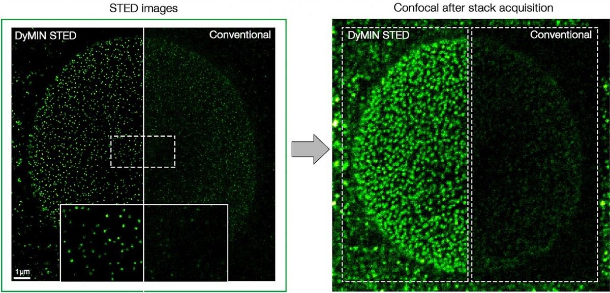
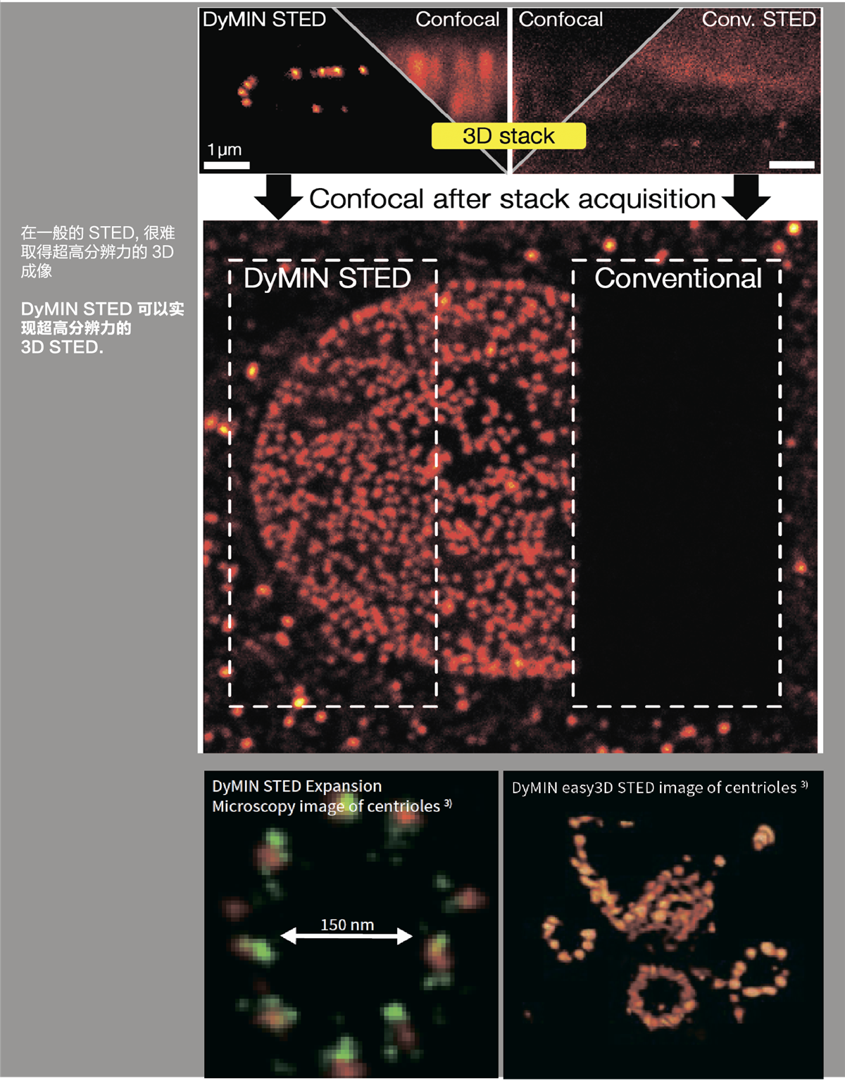
After acquisition with DyMIN STED significantly lower bleaching is observed. Nuclear pore complex protein (nup153) labeled with Oregon Green 488; Excitation: 488 nm, STED: 595 nm.
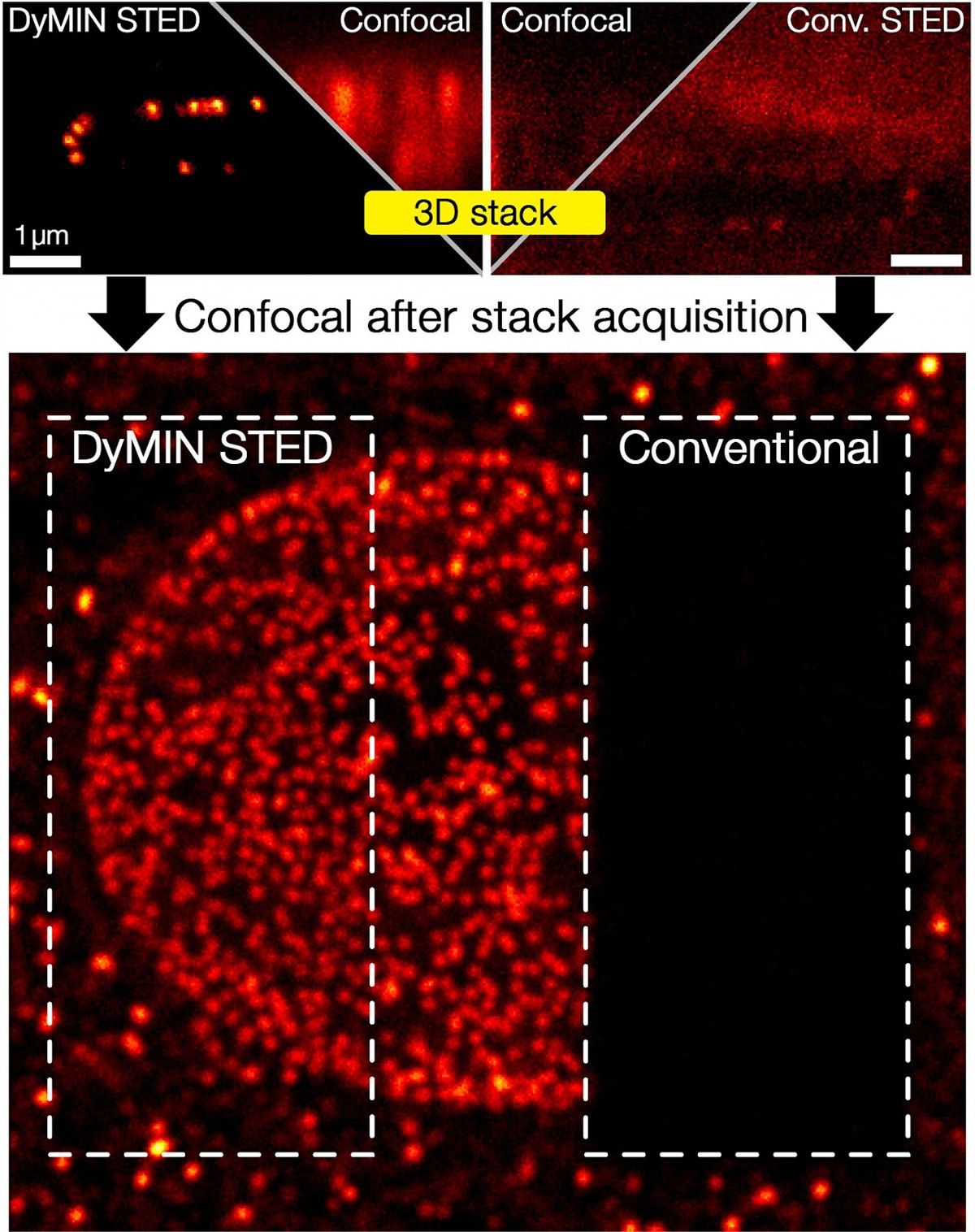
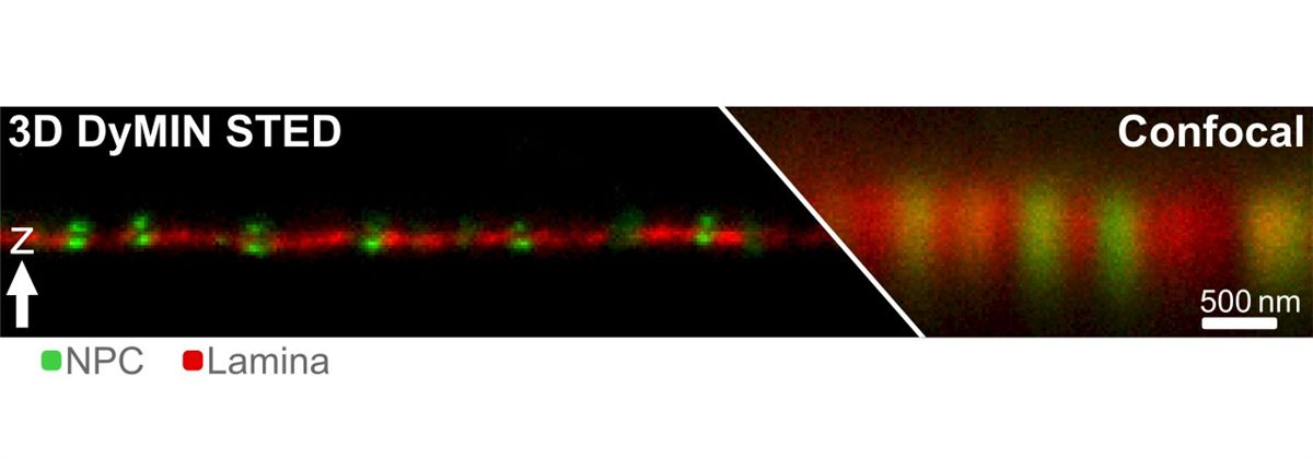
A 3D image stack of the mammalian nucleus was recorded with easy3D DyMIN STED and conventional STED microscopy. Shown are xz sections of the stack and a confocal image after the stack-acqusition, highlighting the remarkable bleaching reduction of DyMIN STED. Note that DyMIN STED enabled us to acquire the complete stack with superior resolution and signal. Shown are Vero cells labelled with antibodies against nuclear pore complex protein (nup153).